35-Point Eye Test
Our Optometry team have created a Unique Operating System (UOS) that details a quality standard for our consultations – we call it our 35 Point Eye Test.
The important first part of an eye examination (any consultation for that matter) is to create a Case History. Your optometrist will collect information regarding the history of your eyes and vision as well as any general health issues, medication, allergies and family history which can impact on risk factors for the health of your eyes and your vision.
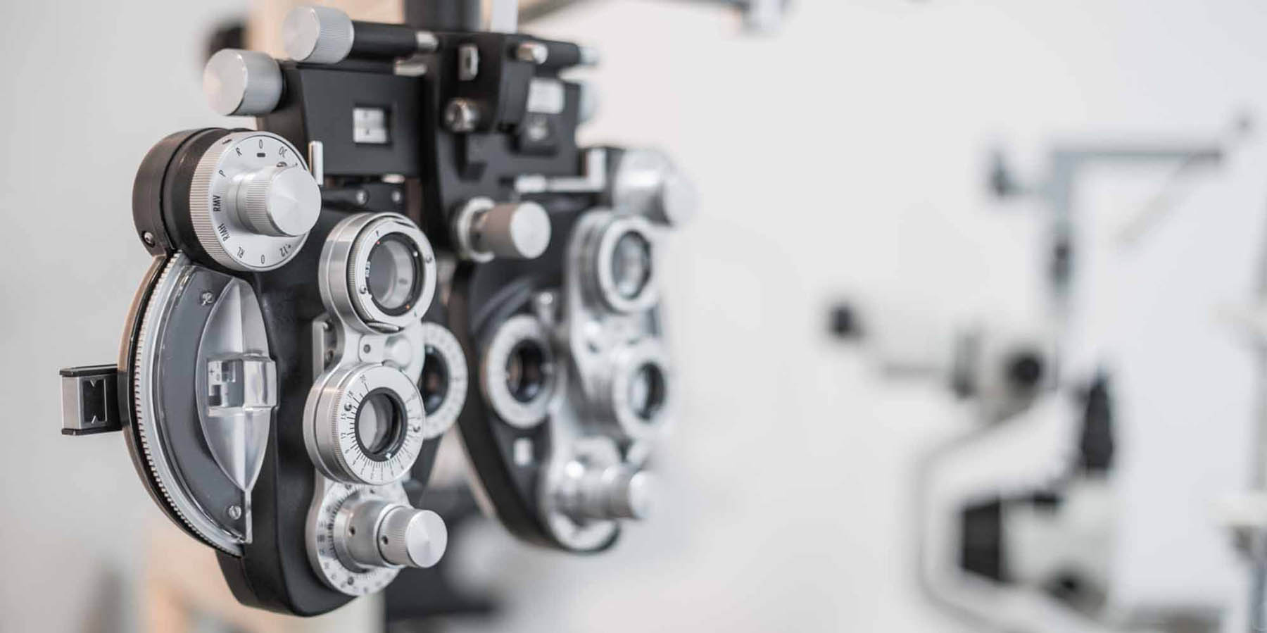
Your vision in the distance without spectacles will be measured. The optometrist will then work out your distance prescription to see if any lenses will help you see more clearly. You will be asked which lenses are clearest. Once this has been determined the optometrist will also measure your prescription for near work, including for reading and the computer if you use one.
After the spectacle prescription (if any) has been determined the optometrist will examine the health of your eyes. Local anaesthetic and orange drops will be used so that the optometrist can measure the pressure in your eyes and have a look at the front surface of your eyes. An increase in the pressure in your eyes makes glaucoma more likely. The optometrist will also use a slit lamp to shine some light on the front of your eyes to examine the structures there including the cornea, conjunctiva, iris and lens.
It is also important to check the health of the inside of the back of your eyes; including you macula, retina and optic nerve. To do this your optometrist needs a good view in through your pupils so they may need to be dilated. If your pupils are dilated you will notice things are fuzzy and glary for an hour or so after your test so we recommend you don’t drive whilst your eyes are affected. Photographs will also be taken of your retina so that it can be documented and any future changes noted.
Lastly, your optometrist will explain the outcome of your eye examination and let you know if eye exercises, glasses, contact lenses or further examination such as visual field tests or OCT examinations are needed and why.
- Case History
- Motility
- Cover test
- NPC
- Pupils
- PD
- UVA (D&N)
- VA with specs
- Phorias with own specs
- Retinoscopy
- Subjective Refraction
- VA R&L
35 Eye Checks
- Binocular Balance
- Phorias with new refraction
- Near Add
- Near Phoria
- Trial Frame
- Slit Lamp
- Lids and lashes
- External adnexae
- External adnexae
- Conjunctiva
- Corneas
- AC/angles
- Lens
- Fundoscopy
- C/D ratio
- Disc margins
- Macula
- Vessels
- Periphery
- Photos
- Diagnosis
- Management
- Advice